Unveiling the arrangement of nanocellulose using scanning electron diffraction
- Mathias Nero
- PhD student,
- Stockholm University
- Co-author(s): Tom Willhammar
- Supervisor (PhD-students/postdocs): Tom Willhammar
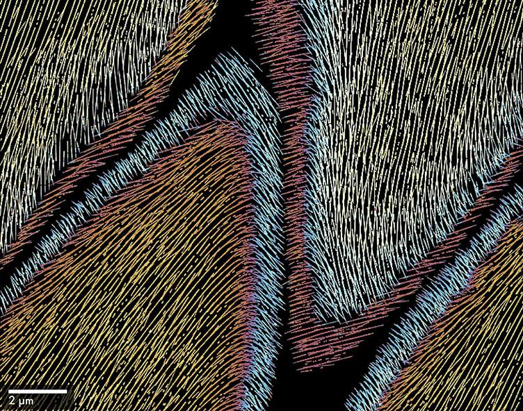
- Cellulose, the primary structural component of wood, exhibits a complex and organized architecture spanning from the nanoscale to the macroscale. A comprehensive understanding of this native hierarchical structure is essential for optimizing the use of cellulose nanofibrils in advanced biomaterials. In this study, we use Scanning Electron Diffraction (SED), a cutting-edge electron microscopy technique, to investigate the arrangement of cellulose within transparent wood, a hybrid material with retained wood structure. SED scans a near-parallel electron beam across the sample in a raster pattern, capturing diffraction patterns at each beam position with a spatial resolution down to 15 nm. By analyzing these diffraction patterns, we revealed that cellulose nanofibrils are highly aligned, from nearly parallel to the cell elongation in the inner part of the cell wall to gradually shifting toward a helical pattern in the outer region. This preserved cellulose structure enhances the mechanical properties of transparent wood. Our findings demonstrate the potential of SED to advance the understanding and development of cellulose-based materials significantly. Figure 1. Virtual orientation map revealing the continuous alignment and orientation of cellulose nanofibrils within longitudinally sectioned transparent wood obtained from a region where two wood cells meet captured with a step size of 100 nm. Black indicates regions lacking cellulose (e.g., middle lamella).
- TIme of presentation: 11:00